When Were Linac Machines First Utilised For Radiosurgery?
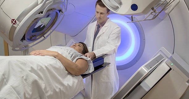
In a radiotherapy centre, there are generally two types of precise radiosurgery technologies available that are tailored to treat particular conditions.
The first, oldest and best known is the Gamma Knife, Lars Leksell’s stereotactic radiosurgery technique that uses multiple smaller beams precisely aimed to destroy tumours and lesions without any harm to surrounding tissue, in keeping with his philosophy that no tool used on the brain can be too precise.
However, Gamma Knife remains one of the leading treatments in precision radiotherapy and has been since its first use Sophiahemmet in 1968 and had been experimented with since at least 1949, the path to the use of Linac machines has been somewhat bumpier, and took longer for its use in radiosurgery to be truly appreciated.
Parallel Developments
The first linear particle accelerator (or Linac for short) was proposed in 1924 by Gustaf Ising and built four years later by Rolf Wideroe, but it was not until after the Second World War that the high-frequency oscillators needed to make Linacs useful for X-rays and radiotherapy possible.
The first Linac installed for clinical purposes was in Hammersmith Hospital, London in 1952, primarily used for conventional fractionation.
Fractionation is, in many respects, the opposite of radiosurgery, and is the use of multiple sessions of radiotherapy over the course of multiple weeks, which maximises the effects of the radiation on tumours whilst protecting healthy cells as much as possible.
In the absence of precision caused by difficulties in keeping Linac beams focused in the early 1950s, this was the best way to take advantage of the benefits of radiotherapy with the tools available, back when radiosurgery was limited to the brain through the Gamma Knife process.
Once Gamma Knife became widely available starting in the 1970s, it became the front-line treatment for brain tumours and other similar conditions in the brain, whilst Linac was primarily used fractionally for long-term radiotherapy, combined therapies with chemotherapy or for palliative purposes.
By the 1980s, however, as technology matured and more was learned about the role of radiosurgery in various treatment pathways, neurosurgeons started to look into the potential for using Linac machines to help treat certain types of epilepsy or arteriovenous malformations.
The first step towards this was the work of J. Barcia-Salorio, a neurosurgeon from Spain who was the lead writer of a 1982 paper suggesting that a potential alternative to invasive surgery would be the use of photon radiosurgery, either using radiation generated from cobalt or using a Linac.
This effectively meant starting from scratch when it came to developing an effective, accurate radiosurgical system, aided by advances in computerised tomography not available back when Lars Leksell was working on the Gamma Knife but had just started to mature in the 1980s.
The first system that took Dr Barcia-Salorio’s conceptual ideas for a Linac radiosurgery system was in 1984 in a paper by O. Betti and V. Derechinsky, both based in Buenos Aires, Argentina
Their system used a frame similar to Dr Leksell’s, albeit using Talairach space rather than Dr Leksell’s own coordinate system, and combined that with intense cross-firing Linac photon beams that converge at the same point to provide an intense dose of targeted radiation without affecting the tissue in the way.
It highlighted that Linac technology had advanced to the point that it was precise enough to at least consider using it as a versatile alternative to the Gamma Knife, and after the 1984 paper, a number of radiosurgery experts started to look into solving the remaining issues surrounding Linac’s precision.
The big leap forward came with the work of Ken Winston and Wendell Lutz, who refined and improved the stereotactic positioning apparatus used and developed a method to measure component accuracy that was previously unavailable but served to benefit radiosurgery as a whole.
The first ever patient to be treated with a Linac-based radiosurgery machine was at Brigham and Women’s Hospital in Boston, part of Harvard Medical School in February 1986.
From there, Linacs have evolved further and become highly capable for both fractionalised radiotherapy and radiosurgery, both with a single focused beam or using the stereotactic process.
Typically, the difference is versatility, as Linacs typically require modification in order to be effective for radiosurgery, whilst Gamma Knife was designed from the start to be used for radiosurgery.
However, image guidance tools, N-localisers and advanced treatment planning tools have helped to make Linac machines more suited for radiosurgery and put the patient’s needs at the centre of any potential treatment pathway choice made by a radiographer.


