Some patients who need to undergo radiotherapy will be told about linear accelerators, which are also known as linacs.
These machines provide external beam radiation to treat cancer patients, as they deliver targeted rays into the patient’s tumour.
How do linac machines work?
Linac-based radiotherapy works by using high-energy X-Rays. They speed up electrons and conform them to the tumour’s shape and size. These then collide with a heavy metal target, which produces the X-Rays.
When these are aimed at the cancer, they destroy the invasive cells but, as they are the right size and shape and specifically targeted, they do not harm the healthy ones.
Therefore, it is effective at keeping surrounding tissue safe, which is why it is a popular choice of radiotherapy treatment.
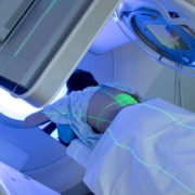
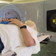
It can be used on all areas of the body, with the patient lying on a moveable couch. Lasers make sure they are in the correct position, so the radiation is beamed at the right area exactly.
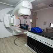
The X-Rays come out of an area of the machine called a gantry, and this can be rotated around the couch, so that the X-Rays can be delivered to the tumour from a number of angles, helping to fully attack the cancerous cells.
The amount of time the patient is in the accelerator will be down to their oncologist, who will also determine the dose they need. Once they have prescribed the treatments, the treatments are usually carried out by radiation therapists.
At the same time, the number of treatments they require will depend on the severity of their cancer and how much radiation therapy it requires. Typically, patients have their treatment spread over a course of a few weeks.
Although patients will hear a buzzing noise while the accelerator is on, they will not feel any pain during the treatment.
What is it used to treat?
Linac-based radiation therapy is commonly used on malignant tumours, as it can deliver high doses to the cancerous cells. It is also used for benign lesions, with electrons being beams being used on superficial lesions.
They can also be used for Total Skin Electron Therapy (TSET), which treats cutaneous T-cell lymphoma. This condition, a form of non-Hodgkin lymphoma, involves lymphomas appearing on the skin all over the body.
Linac is also used in Total Body Irradiation (TBI), which provides the body with a low dose of immunosuppression, and is typically used during bone marrow transplantation procedures.
Patients undergoing prone breast irradiation may also need linac radiation, as this helps reduce radiation toxicity to the lung and heart by letting the patient lie on their front for the treatment.
Prostate treatment with a hydrogel space also requires a linear accelerator technology. This procedure involves injecting liquid Hydrogel between the prostate and the rectum so there is extra space in this area, reducing the toxicity from the radiation.
Side effects of linac radiotherapy?
Like all radiotherapy, linac-based treatments come with side effects.
These can include irritated, dry, red or itchy skin at the site of the radiation; loss of appetite or difficulty swallowing; bladder irritation and urinary problems; changes in bowel habits; headaches and fatigue; and low immunity due to it affecting bone marrow function.
What other types of treatments are available?
While linear accelerator radiation treatments are very effective at destroying cancer cells, there are other procedures available.
For instance, internal radiation therapy might be a better option for some patients, such as those with head, neck, breast, cervix, prostate and eye cancers. As these can be harder to treat in a linac machine, oncologists could prescribe internal radiation therapy, such as brachytherapy, instead.
Alternatively, stereotactic radiotherapy or radiosurgery is a better option for those with brain tumours, as this involves higher doses of radiation in a reduced number of sessions.
As well as brain cancers, it can be used to treat other small tumours with millimetric accuracy, whether they are primary or secondary cancers.
Doctors might also suggest Arc Therapy or Rapid Arc Therapy (VMAT) instead, as this is another effective treatment for cancer.
This involves a radiation beam moving around the tumour in an arc, automatically changing the dose as it moves. Subsequently, this ensures healthy cells receive as minimal an amount of radiation exposure as possible.
Treatment times for this form of therapy is typically only a few minutes, which means patients can be treated more quickly and avoid nasty side effects.