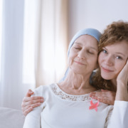
What Should You Expect During Brachytherapy Treatments?
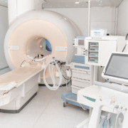
There are several options available for treating tumours, but whilst the majority of radiotherapy treatments use an external source of radiation, brachytherapy implants a radiation source inside the body close to a tumour to destroy it from within.
It is most commonly associated with treatment for prostate or cervical cancer, although it has been used as part of breast, oesophageal, eye and skin cancers, as well as exceptionally rarely during the treatment of brain tumours.
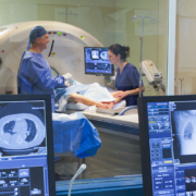
Because a lot of people who receive treatment for cancer do so through some form of external beam radiotherapy (EBT), it can sometimes be difficult to know what to expect during internal brachytherapy.
Here are answers to some of the most common questions people have about internal radiotherapy.
Why Is Brachytherapy Used?
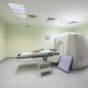
Brachytherapy is a highly accurate and precise set of treatments where radioactive material is placed in the body close to or directly inside the tumour in order to destroy it from within. This can either be temporary using a catheter or using radioactive seeds implanted next to the tumour.
These are known, respectively, as high-dose rate and low-dose rate brachytherapy.
As a primary cancer treatment, brachytherapy is primarily used to allow for higher doses of radiation to be applied to a target area without damaging healthy nearby tissue.
The principle is similar to the highly accurate external Gamma Knife treatment, although it can be applied in other places in the body besides the brain.
It can also be used as part of a combination treatment with external beam radiotherapy or surgery, although it is rare for brachytherapy to be combined with chemotherapy.
Brachytherapy is over 120 years old, and was one of the first versatile types of radiotherapy treatment, and is a proven, safe and effective treatment for a multitude of different types of cancer.
How Long Does Treatment Take?
One advantage of brachytherapy is that it does not require as many sessions as conventional external beam radiotherapy.
High-dose rate brachytherapy typically takes only one or two sessions because a much higher dose of radiation can be used per session, although this can vary depending on your treatment plan. Each session typically lasts no more than 20 minutes from where the radioactive material is in position.
Meanwhile, low-dose rate brachytherapy is typically implanted in a single procedure that takes less than a day but gives off radiation for a few months.
In most cases, you can go home after treatment, although there are some cases where it might be safer to remain for observation for a few nights.
As with other types of radiotherapy treatment, however, you will need someone to take you to and from the hospital due to the anaesthetic used for treatment.
Radioactivity After Treatment
One question that is sometimes asked regarding brachytherapy regards radioactivity following treatment, and whether any precautions need to be made before and after treatment.
The answer depends on the type of treatment, as you are only radioactive whilst a source of radiation is inside your body.
You will be treated in a room on your own and may not be able to have visitors if you need to stay as an inpatient, depending on the types of treatment you had.
If you have radioactive seeds implanted, you will be radioactive, although only close to the treatment area.
You are typically safe to be around most people, but you may be advised to avoid holding children or being in close contact with pregnant women as a precaution. They can remain in the same room as you, but hugging or holding them for more than a few minutes may need to be avoided.
Whilst the radioactive seeds will remain in your body forever, the radiation itself will fade after a few weeks or months. As it does, you will no longer need to be careful with close contact.
Are There Any More Precautions To Take After Treatment?
You will receive medications to help ease any symptoms and receive a card with contact details and an explanation of the treatment you have had.
For the first few days after treatment, the area where you had treatment may feel sore, and you will need to avoid intense exercise and heavy lifting for at least three days.
Make sure to stay hydrated, eat a balanced diet and take any pain medication that you have been prescribed.
The symptoms will get easier after the first few weeks, and you will have regular follow-up sessions to ensure that if there are any unexpected issues that they can be addressed by your doctor and multidisciplinary team.