Study Supports New Brain Tumour Radiotherapy Approach
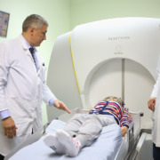
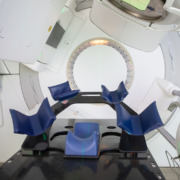
Stereotactic radiotherapy is one of the most advanced forms of radiotherapy. It is designed to achieve the delivery of very concentrated and precise beams of radiation to tumours while minimising the irradiation of any surrounding tissue, which makes it invaluable when tackling tumours and cancerous cells in or adjacent to the brain.
If you are seeking private radiotherapy, your primary aim should be to get the best possible treatment, perhaps something not available via your country’s state health system, using the most advanced and appropriate form of radiotherapy.
What Is Stereotactic Radiotherapy?
Not every form of cancer needs stereotactic radiotherapy, but brain metastases certainly do. Metastatic cancer is a form of cancer in which the disease has spread from its original site to other parts of the body. That could mean brain cancer spreading to other parts of the brain or elsewhere in the body, or another kind of cancer spreading to the brain.
In either case, this is among the most serious forms of cancer anyone can suffer from and significant intervention will be required. The two principal means of treating brain cancer are through surgery to remove part or all of a tumour and stereotactic radiotherapy to shrink it.
Surgery vs. Radiosurgery: A New Approach & Key Findings
These are commonly used in combination in the course of treatment, which means studies to find ways of delivering this combination in ever more effective ways are a very active area of research around the globe.
In a particularly notable development revealed this month, research in the US has indicated that a new method of combining stereotactic radiosurgery (SRT) with surgery is not only safe, but displays enhanced outcomes compared to the alternative approach.
The randomised clinical trial by the Eastern Cooperative Oncology Group, published in the Journal of American Medical Association, set out to establish the safety and effectiveness of using SRT on patients before operations to remove resectable brain metastases (cancerous cells that can be removed surgically), with the purpose being to compare outcomes with the alternative of carrying out SRT after surgery.
What the study concluded was that the pre-operative SRT was just as safe for patients as post-operative treatment. In addition, the pre-operative treatment produced two positive benefits, with more treatments being completed and the time taken to finish treatment being reduced.
Among those in the cohort who had pre-operative SRT, 88 per cent completed both stages of treatment, compared to only 73 per cent in the post-operative SRT patients, while the median gap between the two treatments was only six days for the pre-operative SRT group, compared with 22 for the other group.
Implications for Private Cancer Treatment
As with any study of this type, it will require peer review and further research will be needed to corroborate the findings. However, if it is confirmed that pre-operative SRT is the better option for patients with resectable metastases, it may become the standard procedure.
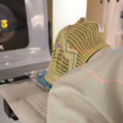
This kind of cutting-edge research is meaningful because the technology to carry out such work is now so advanced. SRT has been developed with increasing precision over the years, with tools like the Gamma Knife, first pioneered by Swedish scientist Lars Leksell in the 1960s, being enhanced with a second version in 1975 and further improved since.
Managing Side Effects of SRT
Both SRT and surgery will require significant recovery time and will come with a range of side effects. In the case of any radiotherapy treatment, common symptoms may include hair loss, nausea, vomiting, stomach troubles and loss of appetite, tiredness, skin problems and loss of libido.
It is worth noting, however, that not all of these symptoms will occur and some kinds of radiotherapy can produce certain side-effects more than others.
For example, blood-related issues caused by a reduction in blood cell production are commonly caused by external beam radiotherapy and internal isotope radiotherapy, because these impact the bone marrow where blood cells are produced. This is not, therefore, a common symptom of SRT.
What To Expect From Amethyst
The purpose of the study in the US was to find the best way to combine radiotherapy and surgery. In your own case, the brain cancer may not be metastatic, but the best treatment may still involve both SRT and surgery.
Where this is not the case, it will be because the cancer exists in the form of a tumour in a part of the brain that cannot be safely accessed in physical surgery. In this instance, SRT will have an even more important role to play.
What you can be sure of is that the treatment we offer will use the very best methods and knowledge at our disposal.
Learn more about our advanced radiotherapy treatments for brain cancer on the Amethyst Group website.