How Is Radiotherapy Used To Treat Prostate Cancer?
Prostate cancer is one of the most common forms of cancer affecting men. The most recent EU-wide figures (for 2020) showed it accounted for 23 per cent of cancer diagnoses in men, while data for the UK between 2017 and 2019 put the figure at 28 per cent.
There are some significant demographic variations. For instance, the disease is most likely to affect men in their late 70s, while there are also racial differences, with black men at the greatest risk and Asian men at higher risk than their white counterparts.
New Discovery Highlights Genetic Risk
A further discovery has highlighted how some men may be at a particularly high risk for genetic reasons. The Institute for Cancer Research in London has found that men with mutations in the BRCA1 or BRCA2 genes are at an elevated risk of getting the disease.
This led to the institute calling for changes in UK prostate screening guidelines to ensure that all men aged over 40 with these mutations are given regular screening to increase the chances of an early diagnosis.
The findings were presented at the annual congress of the European Society for Medical Oncology. It marks a shift from the previous findings from the same institute, which had already identified mutations in the BRCA2 gene as a sign of increased risk, with sufficient evidence now available to add mutations in the BRCA1 gene to the high-risk category.
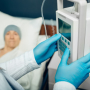
Treating Prostate Cancer
As with any type of cancer, an earlier diagnosis can make treatment easier. However, whether the diagnosis is early or late, radiotherapy will commonly be used.
While earlier detection typically broadens treatment options and improves outcomes, radiotherapy remains a principal treatment for prostate cancer across different stages.
Our private radiotherapy centre can offer different methods of delivering this, in accordance with each patient’s circumstances.
Because prostate cancer is so common, there is a lot of knowledge and treatment about how to tackle it. However, as can be seen above, research is ongoing on a large scale and more is being learned all the time. This means that new means of treating the disease emerge frequently, whether entirely new or a fine-tuning of existing methods.
In the case of radiotherapy, the latter applies, as radiotherapy as a treatment for prostate cancer has a long history. It may be used on its own, or in combination with other treatments such as surgery to remove the tumour or even the whole prostate, as well as chemotherapy.
There are two main methods of treating prostate cancer through radiotherapy, which are external beam radiotherapy and brachytherapy.
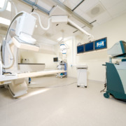
External Beam Therapy
In the first case, this involves beams of radiation being aimed at the affected area using a machine to direct them. The most precise form of external beam radiotherapy is known as stereotactic radiotherapy, which seeks to target very specific, small areas. That is something that can be done when the cancer is very localised, such as after an early diagnosis.
The benefit of stereotactic radiotherapy is that it can concentrate the radiation very closely on a small area to target the cancer effectively while minimising exposure for the surrounding tissue.
For this reason, less concentrated radiotherapy may be more likely to be used in the event of secondary cancer occurring, which spreads beyond the prostate into surrounding areas and therefore warrants the delivery of a wider dose of radiation across a less restricted area.
The Use Of Brachytherapy
The other main treatment method is Brachytherapy. This involves placing a small seed of radioactive material under the skin in or near the affected area, where it will deliver radiotherapy. This means it is delivering this constantly rather than in specific sessions at one of our radiotherapy centres.
In the case of brachytherapy, multiple ‘seeds’ of material, around the size of a grain of rice, are used, with the number varying according to the size of the tumour.
If it proves possible to use radiotherapy alone without resort to surgery, this can produce better outcomes for the patient. Common problems for those who undergo surgery include losing the ability to get and sustain erections and urinary leakage.
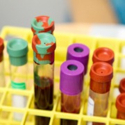
Side Effects Of Radiotherapy
As with radiotherapy for any form of cancer, there are side effects to consider. These commonly include tiredness and fatigue, skin soreness, a loss of appetite, bowel and urinary issues, and sexual side-effects (including erectile dysfunction).
The minimisation of exposure for healthy tissue near affected areas, reducing symptoms, is one of the benefits of stereotactic radiotherapy.
However, the right kind of radiotherapy treatment, along with other treatments, will depend on the circumstances of each patient. What we will always do is ensure that you get the best treatment for your own situation.
Learn more about our advanced radiotherapy treatments for prostate cancer on the Amethyst Group website.