Study Shows MRI Helps Guide Glioblastoma Radiotherapy
Radiotherapy can be used against various forms of cancer, but some of the most cutting-edge and precise uses involve brain tumours.
There are very good reasons for this. Brain tumours vary in their impact, as some are benign and simply need to be shrunk to prevent secondary problems such as pressure on the brain or nerves. Others are cancerous and radiotherapy, alongside chemotherapy and surgery, can extend the life of patients by varying amounts.
With any form of radiotherapy treatment, calibrating the right dose and hitting the right targets is vital. This is especially true when targeting a brain tumour, given the dire consequences of damaging healthy brain tissue nearby, but there is also the general consideration that the less radiation there is, the less severe the side effects.
For this reason, anything that helps increase the precision of the radiotherapy, either in its delivery or in the preparation and assessment of treatment will help ensure it is given in the right places, in the correct doses and that this is linked to the correct supplementary treatment.
The Importance Of Tackling Glioblastomas
In the cases of benign tumours, this can help with the process of enabling the patient to live a normal life. For those with cancerous tumours, the challenge is greater, especially in cases such as glioblastomas, aggressive tumours arising from glial cells that show up in the brain and spinal cord and can, if unchecked, kill sufferers within months.
With the best treatment, however, some glioblastoma patients can live for up to five years, so there are clear benefits to be gained from good treatment. Moreover, as they are the most common form of brain tumour, accounting for 32 per cent of all cases, there is plenty of incentive to develop treatments to improve outcomes for patients.
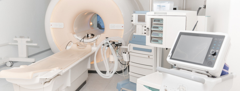
For that reason, new research in the United States has indicated that patients may benefit from a novel new use of MRI scans to help guide radiotherapy on glioblastomas, with the two being delivered simultaneously to help provide a new method of analysis.
MRI, Radiotherapy and Real-Time Data
The study on this approach was carried out by researchers at the Sylvester Comprehensive Cancer Centre in the University of Miami’s Miller School of Medicine. The research was published in the International Journal of Radiation Oncology – Biology – Physics, as well as being presented at a meeting of the American Society for Radiation Oncology.
What this showed was that having daily MRI scans delivered alongside the radiation therapy could help guide radiotherapy treatment more precisely, as well as keep track of developments, which meant oncologists could make adjustments daily to treatment when necessary to deal with the very latest developments and tackle them accordingly.
Known as MRI-linac, the method produced scan results that matched the normal standard MRI scans carried before and after courses of treatment in 74 per cent of cases.
In the other 26 per cent of instances, the MRI-linac system projected tumour growth when in fact it shrank. However, this does mean that, crucially, there were no instances where a tumour increased in size without the MRI-linac predicting this would happen.
Why The System Could Enhance Radiotherapy For Glioblastomas
Consequently, an effective way to utilise the system is to use contrast imaging as a follow-up means of confirming if a tumour is indeed growing, which, the study suggests, will be the case in three-quarters of indicated cases.
Lead author Dr Kaylie Cullison, said: “Our study shows that these daily scans can serve as an early warning sign for potential tumour growth.”
The key finding was that using MRI scans is more effective in spotting developments than standard imaging techniques, helping offer better treatment guidance and therefore paving the way for better treatment. The research team has concluded that this could eventually become the standard approach for using radiotherapy on glioblastomas.
At present, this is not the standard way for treating glioblastomas, with Sylvester Comprehensive Cancer Centre being unique in providing this approach, but that may change over time. However, it adds to an array of new techniques, technological developments and ongoing research that could enhance the care of patients.
Radiotherapy Technology Keeps Progressing
Many of these developments have occurred in specific areas, such as the development of ever more precise gamma knife technology. Invented by Swedish scientist Lars Leksell in 1967, the invention has been further refined, first with the second version in 1975 and several times subsequently as the technology has advanced.
If you need radiotherapy, our staff will guide you through not just what you can expect in a course of treatment, but explain how various developments have enabled patients to enjoy better outcomes as a result of increasingly better technology, diagnostics and understanding of tumour treatments.


